


The Cardiac Valves
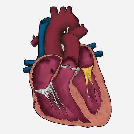
Heart valves prevent the back flow of blood through the cardiac system, maintaining normal unidirectional flow. The four valves of the heart, in the order of blood flowing through them, are named:
-
Tricuspid valve: separates the right atrium from the right ventricle and prevents back-flow into the right atrium during ventricular systole
-
Pulmonary valve: prevents back-flow from the pulmonary artery into the right ventricle during diastole
-
Bicuspid (mitral) valve: prevents back-flow from the left ventricle into the left atrium during systole
-
Aortic Valve: prevents back-flow from the aorta into the left ventricle

The Tricuspid Valve
Axial View
Frontal View
The tricuspid valve is an atrioventricular valve located between the right atrium and the right ventricle. It is composed of three leaflets—anterior, posterior, and septal—anchored by chordae tendineae to papillary muscles in the right ventricle. The valve opens during atrial contraction to allow blood to flow into the ventricle and closes during ventricular contraction to prevent backflow into the right atrium, ensuring efficient one-way circulation on the right side of the heart.
The Pulmonary Valve
Axial View
Frontal View
The pulmonary valve is a semilunar valve located between the right ventricle and the pulmonary trunk. This means it sits anteriorly within the heast cavity. It consists of three crescent-shaped cusps that open during ventricular contraction to allow blood to flow into the pulmonary arteries and close during relaxation to prevent backflow into the right ventricle. The pulmonary valve ensures one-way blood flow from the heart to the lungs and plays a key role in maintaining efficient pulmonary circulation.
The Mitral Valve
Axial View
Frontal View
The mitral valve (also called the **bicuspid valve**) is the atrioventricular valve between the left atrium and the left ventricle. It has two leaflets (an anterior one and a posterior one), which are tethered by chordae tendineae to strong papillary muscles. During ventricular filling, the valve opens to allow oxygenated blood to flow into the left ventricle. During ventricular contraction, it snaps shut to prevent backflow into the left atrium. Because it regulates blood entering the heart’s main pumping chamber, the mitral valve plays a crucial role in generating the high pressures needed to supply the entire body. Damage to the mitral valve often results in atrial fibrillation.
The Aortic Valve
Axial View
Frontal View
The aortic valve is a semilunar valve located between the left ventricle and the aorta, guarding the exit from the heart’s main pumping chamber. It consists of three cusps—the right, left, and non-coronary cusps—each forming a small sinus that helps the valve close smoothly. During ventricular contraction, the valve opens to allow blood to surge into the aorta; when the ventricle relaxes, it closes firmly to prevent backflow. Notably, the coronary arteries arise from the aortic sinuses, making the aortic valve essential not only for systemic circulation but also for supplying blood to the heart itself.


Valves In ECHO Imaging
In cardiology, we use a multitude of different imaging modalities to visualise different parts of the heart. The most easily accesible test we have is the echocardiogram. As we are primarily focussed on ECG interpretation, we suggest visiting our friends at "Medmastery.com" if you would like to learn more about ECHO imaging


Pulmonary valve and Aortic valve
On echocardiography (ECHO), the pulmonary and aortic valves are both semilunar valves but are assessed in different views and pressure settings.
Pulmonary valve:
The pulmonary valve is best seen in the parasternal short-axis and right ventricular outflow tract (RVOT) views. It appears as a thin, trileaflet valve that opens widely during systole as blood is ejected into the pulmonary artery. In diastole, the cusps meet neatly in the center, forming a smooth line of closure. Doppler typically shows low-velocity, laminar systolic flow, consistent with the low-pressure right-sided circulation.
Aortic valve:
The aortic valve is most clearly visualized in the parasternal long-axis and parasternal short-axis views. It is a trileaflet valve with thicker cusps than the pulmonary valve due to higher pressures. In systole, the cusps open symmetrically, creating a characteristic “Mercedes-Benz” appearance in short-axis view; in diastole, they close tightly to prevent regurgitation into the left ventricle. Doppler assessment shows higher systolic velocities than the pulmonary valve, reflecting the high-pressure systemic circulation.
See the ECHO parasternal short axis images below for reference.
TV = Tricuspid valve
RA = Right atrium
IAS = Inter-atrial septum
LA = Left atrium
LV = Left ventricle
RV = Right Ventricle
RVOT = Right ventricular outflow tract
PV = Pulmonary valve
PT = Pulmonary Trunk
NCC = non-coronary cusp (of aortic valve)
RCC = right coronary cusp (of aortic valve)
LCC = Left coronary cusp (of aortic valve


Tricuspid and Mitral Valves
On echocardiography (ECHO), the tricuspid and mitral valves are atrioventricular valves and are best assessed together in the apical four-chamber (A4C) view, which clearly shows their relationship to the atria and ventricles.
Tricuspid valve:
In the apical four-chamber view, the tricuspid valve is seen between the right atrium and right ventricle, positioned slightly more apically than the mitral valve. It has thin, mobile leaflets that open widely during diastole to allow right ventricular filling and coapt during systole to prevent regurgitation into the right atrium. On color Doppler, normal flow is laminar with low velocities, reflecting the low-pressure right heart, while regurgitant flow—if present—directs back into the right atrium.
Mitral valve:
The mitral valve is visualized between the left atrium and left ventricle and appears more basal than the tricuspid valve in the same view. Its anterior and posterior leaflets are thicker and more robust. During systole, the leaflets close firmly with a smooth line of coaptation. Doppler interrogation demonstrates higher inflow velocities than the tricuspid valve, consistent with the higher pressures of the left heart.
In the apical four-chamber view, comparison of these valves highlights the structural and functional differences between the low-pressure right heart and the high-pressure left heart, making it a key view for evaluating atrioventricular valve function.


Other Topics Within This Section
Skip to a particular section


Download Our App Now
For iOS and Android


