


The Cardiac Chambers
The heart is a muscular pump that lies centrally within the chest. It's comprised of four separate chambers:
-
Two atria (the upper chambers) and,
-
Two ventricles (the lower chambers).
The right side of the heart takes deoxygenated blood from the systemic circulation, and pumps it through the lungs to be oxygenated. The left side of the heart then receives the oxygenated blood, and pumps it back through the arterial system to supply the end organs.
Each of the cardiac chambers has its own defining features, depending on its function. The top two chambers are known as atria and are typically thin walled due to the low pressures that they endure. The ventricles are the two lower chambers. Of the ventricles, the left ventricle has the thickest muscular wall, as it is required to push blood all the way through the systemic circulation.


Other Structures Of Note
There are several other notable structures that feature within the heart. As our course is predominantly based around ECG interpretation, we have made sure to point out only the most important and relevant structures. For a more in depth anatomical discussion, please feel free to visit our friends at "Teachmeanatomy.com" and "Khan Academy"

Inter-Atrial Septum
The inter-atrial septum is the thin wall that separates the right atrium from the left atrium of the heart. Its key feature is the fossa ovalis, a shallow depression on the right atrial side that represents the closed foramen ovale, a fetal opening that allowed blood to bypass the lungs before birth. The raised rim around it is called the limbus of the fossa ovalis.
Functionally, the inter-atrial septum prevents mixing of oxygenated and deoxygenated blood between the atria and plays an important role in normal cardiac circulation.

Inter-Ventricular Septum
The interventricular septum is the thick muscular wall that separates the right ventricle from the left ventricle.
It has two main parts: a large muscular portion, which makes up most of the septum and contributes to ventricular contraction, and a small membranous portion near the base of the heart.
The septum plays a key role in efficient pumping by preventing blood from mixing between the ventricles and by helping transmit electrical impulses through the bundle of His as part of the heart’s conduction system.


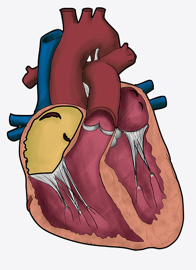
Atrial Appendages
The atrial appendages (also called auricles) are small, ear-shaped muscular outpouchings that extend from each atrium.
The right atrial appendage is broad and triangular, while the left atrial appendage is narrow and elongated. They contain pectinate muscles, which increase contractile strength, and help the atria accommodate changes in blood volume. Clinically, the left atrial appendage is important because it is a common site for blood clot formation in conditions such as atrial fibrillation.


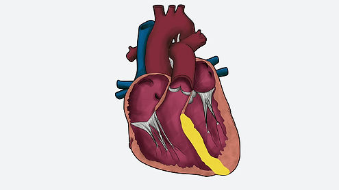
The Great Vessels
The great blood vessels of the heart are the major arteries and veins connected directly to the heart. They transport blood to and from the heart and play a vital role in circulation.
Aorta (highlighted in yellow)
The aorta is the largest artery in the body. It carries oxygen-rich blood from the left ventricle to the rest of the body. After leaving the heart, it arches upward (the aortic arch) and then travels downward, giving off branches that supply the head, arms, and body.
Pulmonary Trunk (Highlighted in blue)
The pulmonary artery carries oxygen-poor blood from the right ventricle to the lungs. It divides into the right and left pulmonary arteries, which transport blood to each lung for oxygenation.
Other Topics Within This Section
Skip to a particular section


Download Our App Now
For iOS and Android


